A tooth does not always have to be extracted: apical microsurgery helps preserve the natural tooth


“Once we extract a tooth, there is no way back. That is why, whenever a tooth can be preserved, we should do so,” says Dr Aleksandra Kārkle, lecturer at the Faculty of Dentistry of Rīga Stradiņš University (RSU), certified endodontist.
This idea of preserving the natural tooth became the basis of her doctoral thesis, Assessment of Bone Regeneration after Apical Microsurgery Using Advanced Platelet-Rich Fibrin, which examined jawbone healing after apical microsurgery and modern ways of evaluating this process.
From clinical practice to scientific research
Her interest in the topic emerged after her residency in endodontics, when Aleksandra Kārkle increasingly encountered patients whose infection had spread beyond the root canal system and affected the surrounding jawbone. “During my studies, relatively little time was devoted to apical microsurgery. I realised that I wanted to explore this field in much greater depth – I trained with international specialists, developed my practical skills and later understood that I also wanted to evaluate treatment outcomes scientifically,” she says.
The central question of the doctoral thesis was not the operation itself, but how the jawbone regenerates afterwards and how this process can be assessed as accurately as possible.
When the tooth does not hurt, but inflammation is already progressing
One reason why patients reach a specialist late is that the inflammation often causes no symptoms. “Most patients feel neither pain nor discomfort. Inflammation is often discovered accidentally during radiological examination, or only when it has already become more extensive and symptoms have appeared,” the doctor explains.
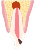
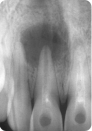
The infection is located in the tooth’s root canal system, while the inflammatory changes develop in the jawbone near the tip of the tooth root. The body tries to fight the infection, and as a result of this process a bone defect gradually forms.
The first choice is always root canal treatment or, if necessary, retreatment. However, this is not possible in every situation. If access to the root canals is difficult, or if previous restorations make retreatment impossible, the tooth can often still be saved with apical microsurgery.
A modern way to preserve the natural tooth
Apical microsurgery is a highly precise surgical procedure during which the doctor accesses the focus of inflammation near the tip of the tooth root through a small opening in the jawbone.
During the operation, inflamed tissue is removed, a small part of the root tip is resected, and the root end is sealed hermetically with a special biomaterial to prevent reinfection. “This is often the last opportunity to preserve the patient’s natural tooth. Today, the procedure is performed at a microsurgical level – using an operating microscope, ultrasonic instruments and microsurgical suturing techniques. This allows the operation to be carried out much more precisely and gently than before,” says Kārkle.
The key question of the study: how does the jawbone regenerate?
The main aim of the doctoral thesis was to evaluate jawbone regeneration after apical microsurgery. The study involved 43 patients who underwent apical microsurgery at the RSU Institute of Stomatology between 2021 and 2024.
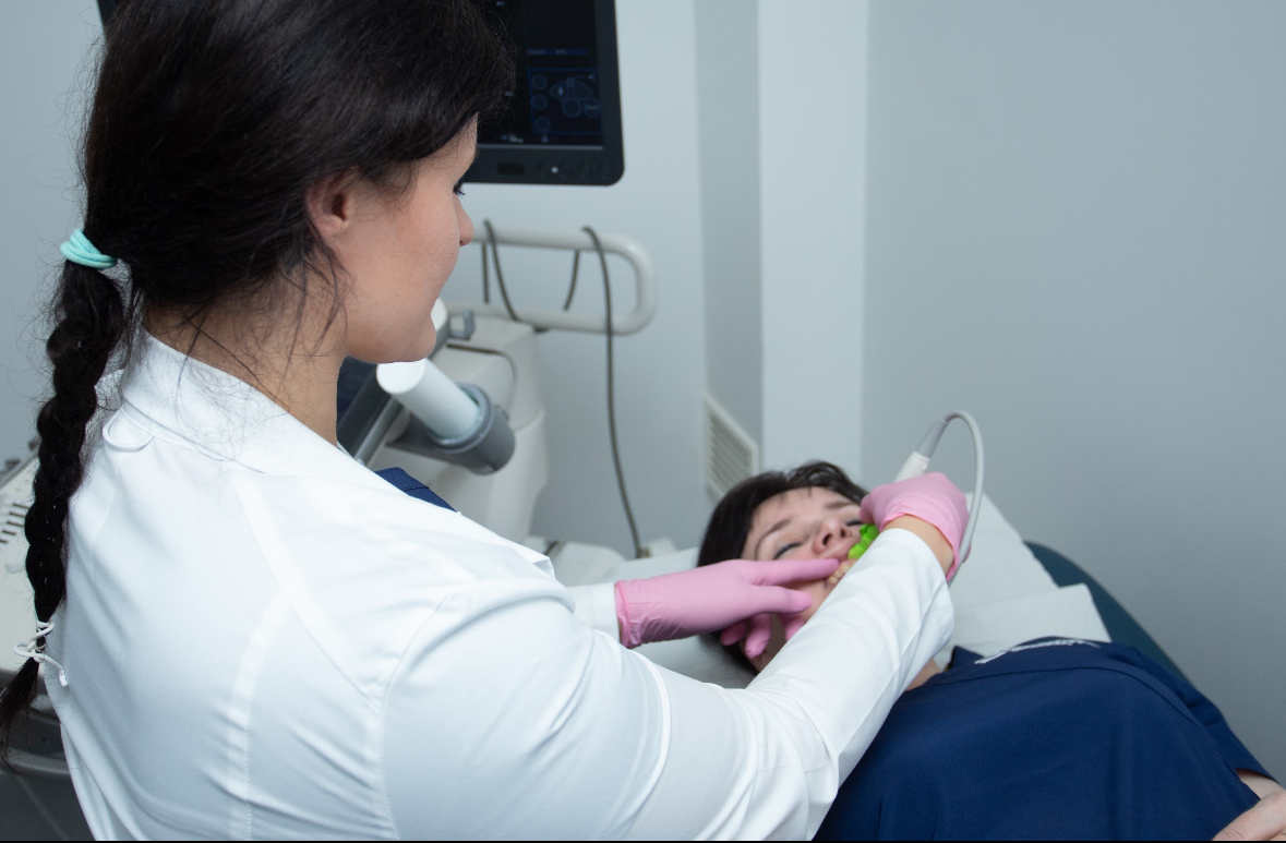
One part of the study compared three imaging methods – periapical radiography, cone-beam computed tomography and ultrasonography – to assess their suitability for diagnosing jawbone defects and monitoring healing.
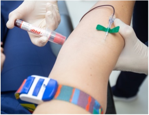
The second part of the study analysed whether bone regeneration could be promoted using A-PRF (advanced platelet-rich fibrin) – an autologous biomaterial obtained from the patient’s own blood shortly before surgery. “A-PRF contains platelets, leukocytes and growth factors. Its potential to promote tissue healing is being studied in many fields of medicine, and we wanted to find out whether it could support jawbone regeneration after apical microsurgery,” says the researcher.
Positive patient experience and future prospects for A-PRF
When jawbone healing was assessed in radiological examinations, no statistically significant advantage of A-PRF over standard treatment was demonstrated. “This does not mean that the biomaterial is not promising. In our study, we were unable to demonstrate a radiological advantage, but A-PRF remains a promising autologous biomaterial whose potential to promote tissue healing is also being actively studied at the biological level. It is possible that some biological processes simply cannot be fully assessed using the radiological methods currently available,” explains Kārkle.
At the same time, patients often described the postoperative period as relatively easy, with only minor discomfort.
Ultrasonography – a promising complementary method
One of the most significant contributions of the doctoral thesis is its evaluation of the use of ultrasonography in endodontics. Until now, radiological methods have mainly been used to assess jawbone healing, but in certain situations ultrasonography can provide valuable additional information.
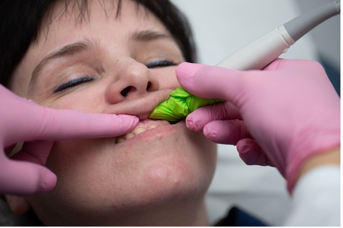
“Ultrasonographic examinations do not use ionising radiation. This means that, in the future, ultrasonography could become an important complementary method for patients for whom it is advisable to reduce radiation exposure, for example during long-term follow-up. It does not replace computed tomography, but in certain cases it can be very useful for assessing jawbone defects and healing,” explains Kārkle.
Preserving the natural tooth is always a priority
The aim of modern endodontics is to preserve the patient’s natural tooth for as long as possible, so that it can continue to fulfil its function. Published data show that the success rates of apical microsurgery reach approximately 90–95% when treatment is performed in line with modern microsurgical principles.
“In medicine, there are no absolute guarantees – there is a prognosis. We can use the most advanced treatment methods and follow all standards, but the healing process is also influenced by the individual characteristics of the body and the nature of the infection. Still, as long as a tooth can be preserved, it is worth using every scientifically grounded opportunity to do so,” emphasises Kārkle.
The doctoral thesis provides new knowledge about jawbone regeneration after apical microsurgery, broadens understanding of the potential of ultrasonography in endodontics and confirms that modern dentistry is placing increasing emphasis on preserving the natural tooth and using gentle treatment methods.
Summary of Aleksandra Kārkle’s doctoral thesis in the RSU e-resource repository
Related news
 Signs of malnutrition and inflammation may help predict complications in microvascular flap surgeryRSU research synapses