New RSU Anatomy Museum Designed to Appeal to Both Medical and Social Sciences Students
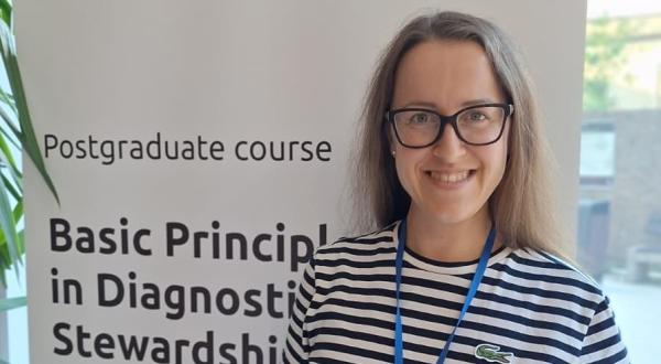
The new Rīga Stradiņš University (RSU) Anatomy Museum was planned to open at the end of 2020. Everything changed due to pandemic, but hopefully the museum will be able to open its doors to visitors in early 2021. Three years ago, Ieva Lībiete undertook to be the head of the museum. At that time, she could not even have imagined that the museum’s construction would be concluded on as historically a significant a year as 2020.
The RSU Anatomy Museum is a structural unit under the RSU Institute of the History of Medicine. It is a new and unique place for medical students, schoolchildren, and the public to learn and conduct research. The museum is located at 9 Kronvalda bulvāris, Riga, on the grounds of the Anatomical Theatre. It will join the Institute of the History of Medicine, which is managed by Professor Juris Salaks, in making a significant contribution to the quality and competitiveness of the study process at RSU as classes in the history of medicine and life sciences will henceforth take place on the museum’s state of the art premises. The Institute of the History of Medicine includes both RSU Museum collections - one on anatomy and the other on the history of the university – thus facilitating the collection, preservation, and interpretation of both the tangible and intangible heritage of medical history. It also makes these collections accessible for research, education, and museology.
Tell us about the history of the museum and how you got involved.
The museum has existed since 1920. When the researchers and students of the newly established Faculty of Medicine needed training materials, they turned to the museum. It was established by the Swedish professor Gaston Backman, who had come to Latvia to become a professor of anatomy and was the Head of the Institute of Anatomy at the University of Latvia. The institute was located on the 2nd floor of the current Anatomical Theatre, and the museum was in its "heart", in the beautiful and monumental former chapel hall of an Orthodox seminary.
From then on, the museum has lived through many changes – while its target audience has changed, and it has since become affiliated with a different institution, it is still located in the chapel hall of the Anatomical Theatre. From the end of the 1980s, the museum was managed by the Pauls Stradiņš Museum of the History of Medicine. However, three years ago, RSU suggested an initiative to take over the collection, create a separate Anatomy Museum and make it available to the general public. Thus, another change in management took place, after which the museum collection returned to the supervision of its historical heir. Around the same time, I was invited to take part in the transition process and create the new exhibition. Prof. Jānis Gardovskis, the RSU Rector at that time, showed great trust in me.
I realised that building a new museum in a new building is a once in a lifetime opportunity.
The biggest challenge was to understand how this historical anatomical collection of bones and organs in formalin could be made useful and interesting to a modern visitor.
You are a doctor by profession, and here all of a sudden, you’re working on a museum…
I started working at the Pauls Stradiņš Museum of the History of Medicine already in the fifth year of my medical studies. The museum had captivated me with its special atmosphere and its aura. After graduating, I underwent a residency in internal medicine because I really thought I would become a doctor. At the same time, I also enrolled in doctoral studies in the history of medicine and began to study the history of psychiatry. I always thought that I would finish my doctoral work, return to medicine and choose a specialisation. In the end, however, I realised that I was more interested in medical history, museums, and working with students than working in a clinic.
I have been involved with medical history for fifteen years. When I started studying, I couldn’t even imagine that the history of medicine would fascinate me more than practical medicine.
What makes it so fascinating?
The history of medicine and working in the museum is exciting and multifaceted. There is a creative and a scientific side, as well as an organisational side. I meet many outstanding, smart and interesting people as part of my job. That is one of the privileges of working in a museum and in the field of medical history. I would that there are no negative sides. And my medical education actually helps me a lot.
What attracted you to creating the museum: the goal, or the process?
This is a museum, not a field of research, but a goal is always important. When you can clearly visualise the result, you have already done most of the work.
I created the contents of the museum with my colleague, the excellent social anthropologist Ilze Sirmā.
Our shared goal was to understand how this unique collection, designed for teaching and research, could be useful and interesting for the modern museum visitor.
What stories does this collection tell? There are stories about both the healthy and the diseased anatomy of the human body, about the origins of the collection itself and about its creators, and sometimes also about the people from whom the exhibits were taken. They are stories about diversity and of changing attitudes towards the human body.
Our goal was to make the museum interesting to visitors and to make the old anatomical collection look beautiful. We wanted the museum to arouse an interest in anatomy, but not cause fear and disgust. It seems as if we have succeeded. The main exhibition, which is only one small part of the museum, has been completed. Now we need to visualise our public programming – our educational programmes, the exhibition plan, cooperation opportunities, etc. The pandemic has, of course, made us adjust our plans and made us think more about different types of digital content for next year.
What is the view on anatomical collections today?
Anatomical collections are ambiguous. On the one hand they are, of course, things – objects of scientific and historical interest, which have accumulated over time for various reasons in museums, universities, etc. On the other hand, these objects have a complicated history, as they have come from people who were once alive, and who were not in a majority of cases asked for their consent to being exhibited in a museum after their death. This is the reality in which we lived in Europe until the middle of the 20th century. This was when ethical and legal questions about the human right to one’s body and the protection of tissues and organs of the deceased began to arise, namely that it is up to individuals to decide what happens to their bodies after death. That is why the collections are ambiguous and the fates of various anatomical collections are so different. Many of them have been destroyed. At the end of the 19th century and the beginning of the 20th century, almost every medical school and medical faculty, every hospital and even most departments had their own anatomical collections to show students, or which had accrued as objects of scientific interest. Around the middle of the 20th century there were changes in how anatomy was taught and in the understanding of the affiliation of deceased tissues. Because of this many collections were simply dismantled and were no longer seen as necessary. In some places, however, they were preserved as the heritage of scientific and medical history.
Even today, universities that have preserved collections like this approach them in one of two ways: some continue to keep the collection, but do not exhibit it publicly, while others choose to exhibit it as an important scientific and historical heritage that can give us information not only about the human body, anatomy and diseases, but also provide a broader insight into various ethical and cultural issues. There are several medical museums in Europe where universities take pride in their collections and make them accessible to the general public, not just to professionals in the field.
Therefore, I am very glad that RSU is one of those universities and has deemed its hundred-year-old collection to be a part of its history, which can reveal a lot of useful and interesting things even to visitors today.
We are trying to do this in a dignified way, and I hope that we succeed.
What did you focus on while creating the exhibition?
Anatomical collections have always fascinated people, and we really wondered what questions visitors would have when they encountered them. One of the main reasons that motivates people to visit an anatomy museum is the desire to see something unusual, something titillating. I remember a visit to the Anatomy Museum when I was at school. It was a great experience because I could see something that was usually only available for the chosen few in their white robes, the doctors and medical students. It was also a sort of a test for whether you would be able to become a doctor or not: would the organs scare you, and would you faint at the mere sight?
We are trying to tell visitors that everything in here is real. Here, you can see things the way that a surgeon or an anatomist sees them. You can see what the human body really looks like, because the anatomical images that we see in anatomy books are, to a certain extent, just an abstraction.
There is an infinite variety of human bodies, and our collection shows countless variations.
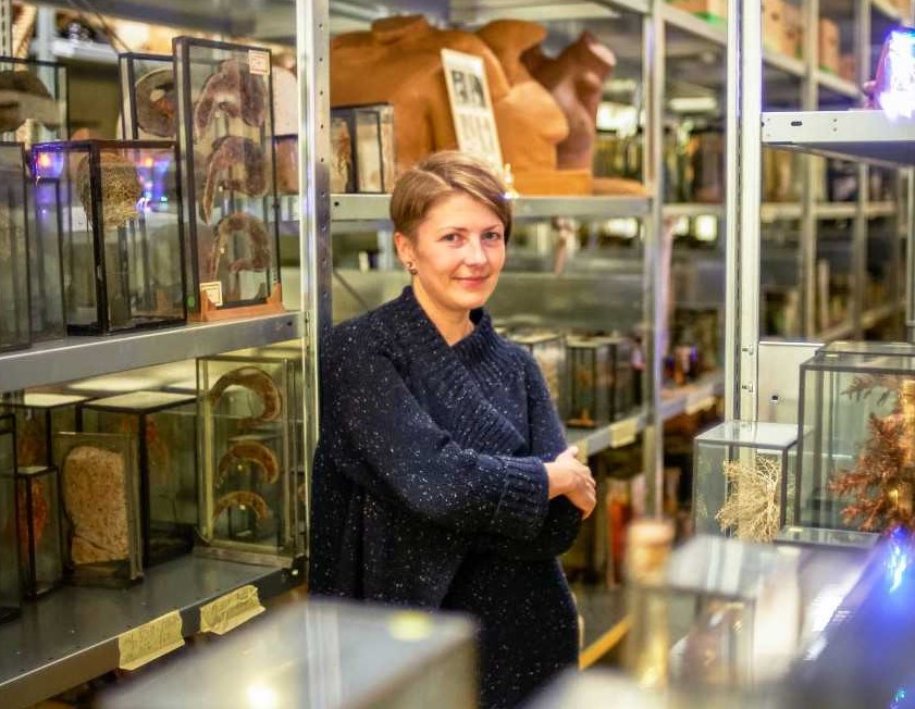
We also tried to make the exhibition answer the question: “How did this object come to be here at the museum?” For example, the museum has a collection of dozens of children’s skulls, which the museum bought in 1920 from doctor Jirgens in Riga. I find this very curious - why did this doctor have eighty children’s skulls at home that he could just sell to the Anatomy Museum? Unfortunately, we haven’t been able to find the answer so far. There are several lists where you can find practitioners in Riga during the 1920s, but Jirgens does not appear on any of them. He may have inherited this collection from one of his teachers who had amassed it for some scientific purpose. The museum also has an anthropological collection - a whole wall covered with skulls. We are often asked why we have so many skulls. Asking this question, you get to hear the story about where they were obtained, what you can learn from them, and why we still keep them today.
When we were just starting to move into the new building, my colleague Diāna Klešnika, the museum’s curator, brought out some teratological specimens from the storage room. The workers who were assembling the exhibition at the time were astounded by what they saw. They were curious about whether they came from Latvians, why they weren’t decomposing, and what liquid they were stored in. The museum’s visitors will have the same questions, so we have tried to answer them all.
There are thousands of specimens stored in the museum’s repositories. We have tried to explain the anatomical, scientific and medical significance of selected objects in the exhibition using digital technologies, animation, and illustrations.
For someone unfamiliar with anatomy, an object in a jar might just seem to be a lump rather than a recognisable part of the body, so we need to provide all exhibits with an explanation. We have also created a downloadable app with 20 different stories. Every museum visitor will be able to use it on their smart device (there will be tablets for rent) to read the medical, anatomical, and historical information that we provide.
Students are crucial to the Anatomical Theatre. How have medical students contributed to creating the museum and how useful will it be for them?
Students are interested in the collections. Some of them have also defended their final theses based on materials from the Anatomy Museum’s collection. There is a fairly large group of students who have volunteered to maintain the collections, like cleaning skulls and bones and to represent the museum in events like Researchers’ Night or Anatomy Night. One of our objectives is definitely to make our collection and the museum lively and student friendly.
In the future, we want to cooperate with lecturers and students from both medicine and the humanities, because the exhibition and the objects in the collection can be researched, explored, and used in various ways, not just from a medical perspective.
We hope to continue our cooperation with lecturers and students from the Art Academy of Latvia.
COVID-19 has now made us discontinue working in person. If it wasn’t for the pandemic, I would certainly already have given lectures in medical history to my students in these rooms – everything you see in the museum can be used in the study process. The anatomical preparations could also be useful in anatomy studies and the teacher can help students interpret them. As I said before, a model or a book are one thing, but seeing an object in real life provides even more understanding. We will see what the future holds for us. It is currently difficult to understand how everything will turn out and how much can still be done in-person.
Groups of schoolchildren frequently visit the Museum of the History of Medicine and the Anatomy Museum. How often do you hear medical students say that it is the museum that inspired them to study medicine?
I work a lot with international students who are not likely to have been influenced by our local museums.
BLOCKQUOTEThe new Anatomy Museum aims to be available to a wide range of visitors. The educational programmes are, however, aimed at schoolchildren from the ninth grade onwards, when anatomy becomes a part of the curriculum.
The Museum of the History of Medicine offers a tour that ranges from traditional medicine to space biology. The Anatomy Museum’s collection, in turn, offers a tour of the human body - its exhibition includes specimens, bones, various organs, castings and so on. The two museums complement each other, their narratives intersect but do not overlap.
One of our goals is to show what medical studies look like in the first year at university and address questions such as: What would it be like if I were to study here? What should I prepare for? When I went to the Anatomical Theatre in the eighth grade, I remember thinking that it was important to see the exhibit since I had decided to become a doctor. But because I come from a family of doctors spanning back two generations, my choice of profession would have been the same regardless of visiting the museum.
At what point do you think that the present becomes history?
It seems to me that history has already begun, because the COVID-19 pandemic will be of interest to medical historians and various social scientists for a long time to come. Just like the black plague, it will be a topic for writing and research for another five hundred years or more.
This is already happening – medical historians and infectologists have become some of the most sought-after experts. When the pandemic had just started, someone called almost every day asking how the history of medicine might help explain what was going on.
What lessons has the history of pandemics taught humanity?
There are medical historians who believe that epidemics follow a certain script.
Charles Rosenberg, a medical historian from Harvard, had a very popular article that was frequently cited during the HIV/AIDS epidemic in the late 1980s. In the article he stressed that all global epidemics so far, and possibly also those in the future, would follow the same scenario in four acts. He based his theory on Albert Camus’ novel The Plague. The first act, or stage, is denial, or gradual recognition. This is when people notice all sorts of small, unfamiliar things that are out of the ordinary, but try to ignore them because they are so unexpected. The Plague begins with Dr. Bernard Rieux leaving his clinic and stepping on a dead rat, which he just pushes to the side with his foot. Rosenberg develops the idea that the same is true of all epidemics – when the first cases arise, people try to ignore them. If a doctor tries to report it, the news does not get attention on a governmental level, because recognising an epidemic would dramatically change normal life. If we look at what originally happened in China at the beginning of the COVID-19 pandemic, it turned out that doctors had already reported it, but that this had gone ignored.
The first act therefore plays out until there are so many victims that the pandemic cannot be denied. And thus, the second act begins – the search for an explanation and, interestingly, for culprits. Who is to blame? This is probably neither the first, nor the last epidemic in human history that China has been blamed for, as many diseases have historically spread from this region. In the Middle Ages, Jews were blamed for spreading the plague. I recently saw a story on Latvian television that Lithuanians were bringing COVID-19 into Latvia across the border. To look for, and find an external culprit is a classic story in the history of epidemics.
The third act, as described by Rosenberg, is responding to the epidemic. The authorities try to bring order by using the resources at their disposal. In Rosenberg’s scenario, these responses are sometimes as devastating as the epidemic itself. This is followed by the final phase of reconciliation and closure, in which everyone is forced to realise what really has happened and how it has changed the world.
The idea that history repeats itself is a captivating dramatic way to see epidemics. But every epidemic is different, of course. We cannot say that we have already been there. All the more so because this is the first disease that has been so widely researched in the history of the world.
Have we learned anything from other epidemics and pandemics, like the Spanish flu? Of course! History has provided an understanding that introducing social distancing measures is an important tool to limit the outbreak, which in turn allows for health care systems to stay open.
The fact that some people do not want to wear face masks today also has historic precedent. People protested against restrictions they did not understand, or believe in during the Spanish flu as well.
Rosenberg would place this type of protests in the third act of his epidemic drama.
When masks were issued in the United States at the time of the Spanish flu, there was a number of people who formed an "anti-mask" league to protest against the use of face masks, believing that their constitutional right to breathe freely was being violated. Human behaviour seems very predictable! I don’t know if there is anything that we can learn from history directly, but it certainly is interesting to know about these events.
What are your expectations, both in regard to the museum and healthcare in general in the new year?
We will certainly try to open the museum up to the public in 2021. Everyone has already been waiting so long for the promised museum. And we, the creators, really want to see visitors’ reactions and feedback. It is clear that for most of the year we will only be available to individual visitors and members of the same household. In summer, if tourism recovers even a little bit, we will welcome tourist groups. Given the current situation, we are aware that groups of schoolchildren will not be crowding the museum next year. We are therefore working on a digital version of the museum’s educational programming, which we hope to be able to offer students already in the spring semester. 2021 will be a virtual and remote year with individual, remote, and masked in-person meetings. Safety comes first!
Information about the Anatomy Museum
- The Anatomy Museum includes a historical anatomical collection created in the first half of the 20th century;
- The museum’s collection is included in the National Museum Collection, which means that it is recognised as a treasure of national significance and is under state guardianship;
- The museum is located in a renovated building specifically adapted to the museum’s needs, in the former stables in the courtyard of the Anatomical Theatre;
- The building has been completely rebuilt and two outbuildings as well as rooms for underground exhibition have been added;
- The museum is completely funded by RSU;
- Curators: Ieva Lībiete and Ilze Sirmā (RSU);
- Design and realisation: Dd Studio;
- The multimedia content of the exhibition was created in collaboration with Anatomy Next;
- Building idea and sketch: Arvīds Līkops (RSU);
- Project by: SIA Delta Construction/ SIA Nams;
- Construction: SIA Velve;
- Parts of two feature films have been filmed on museum premises;
- The museum has published a catalogue about its collection of tattooed skin specimens, Ādas (Skins);
- The museum will open in 2021 when the epidemiological situation in the country improves.