RSU Scientists’ Latest Discovery Explains Why It’s Important to Monitor Blood Calcium Levels
The parathyroid (PT) glands are four small glands in the soft tissue of the neck whose main role is to regulate blood calcium levels. If the level increases, a person can suffer from reduced bone density and fractures, increased formation of urinary tract stones, chronic fatigue, and loss of concentration.
More than 100 patients a year in Latvia are diagnosed with various types of surgically treatable parathyroid pathologies, including benign and malignant tumours. One of the keys to treating this successfully is to improve diagnostic efficiency. At the end of a year-long project that was carried out by radiologists and surgeons from Rīga Stradiņš University (RSU), in collaboration with specialists in endocrinology and pathology,
innovative methods have been found to be able to identify the location and type of damaged parathyroid cells more accurately, which will result in a more precise surgical approach and improved treatment.
The project leaders are radiologist and Associate Professor Maija Radziņa, senior researcher at the RSU Radiology Scientific Laboratory and endocrine surgeon Associate Professor Zenons Narbuts. Here, they tell us more about the project. It was implemented within the framework of the Fundamental and Applied Research Programme and involved scientists from the RSU Department of Radiology, the RSU Radiology Scientific Laboratory, the RSU Faculty of Medicine, the Department of Surgery, as well as medical specialists from Pauls Stradiņš Clinical University Hospital (PSCUH) clinics, radiologist Dr. S. Pavlovičs, surgical resident Dr. E. Tauvėna, medical student M. Ratniece.
PT glands are four small glands in the soft tissues of the neck, behind the thyroid gland. They produce parathyroid hormone, which is involved in calcium metabolism.
As in other organs, both benign and malignant tumours of the parathyroid gland can occur, which in turn can lead to the increased levels of parathyroid hormone in the blood – hyperparathyroidism, and subsequent increased level of calcium in the blood or hypercalcaemia.
Primary hyperparathyroidism is the most common PT-related pathology. It affects 0.1-0.3% of the population and is the most common cause of hypercalcaemia. This is most common in one PT adenoma - a benign tumour.
Primary hyperparathyroidism can cause significant changes in the body due to the accumulation of excessive calcium in the tissues (such as kidney stones) and changes in bone density - osteoporosis, and can affect the body's function at the cellular level (calcium channels).
Secondary and tertiary PT can increase with changes in the body's metabolism - vitamin D deficiency and chronic kidney disease, incl. with renal replacement therapy.
Ultrasound (US) is the most commonly used diagnostic method for imaging parathyroid gland pathologies, based on its relatively low cost, availability, and absence of ionizing radiation. US plays an important role in detecting differences among PT pathologies. Usually, secondary PT pathology is treated with medication.
One of the treatment methods for primary and secondary PT tumours is surgery, therefore precise localisation before surgery is necessary to enable adequate planning of the surgical approach and effective treatment.
Innovative methods such as US elastography and contrast ultrasonography (CEUS) are increasingly being used to improve the accuracy of conventional ultrasonography.
Latvian scientists have explored the possibilities of these new diagnostic methods in characterising EC formations using tissue density measurement (elastography) and contrast ultrasonography (CEUS)
where the patient is administered with a contrast agent intravenously.
Contrast-enhanced ultrasound has the significant effect of expelling air through the lungs, making it safe for use in all age groups, including pregnant women, children and people with chronic kidney disease.
Within the framework of the study, scientists were able to determine the type of PT pathology with greater accuracy, and these methods play an important role in the choice of further treatment and surgical approach, therefore active research is planned in the future.
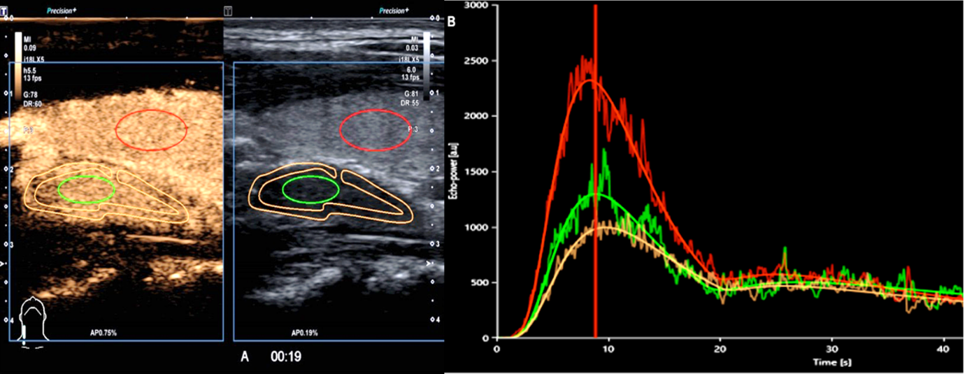 Contrast ultrasonography for parathyroid adenoma
Scientists call on all citizens to pay attention to the state of their health, to the levels of calcium and parathyroid hormones in their blood, and in case of any changes, to approach specialists to help them carry out appropriate modern examinations and initiate treatment in time.
The project is being carried out as part of the LZA grant Izp-2020/2-0297 Multiparametric ultrasound correlation with morphology in patients with primary hyperparathyroidism.